
Presentation
Tripped and fell down steps, landed on left foot with a twisting motion. Barely weight-bearing. Tenderness to palpation over the left dorsal foot and left lateral malleolus. Reduced range of motion with dorsiflexion/plantar flexion and internal/external rotation of the left foot.
Patient Data


Osteochondral fracture of the medial talar dome with displacement of the fracture fragment into the anterior joint space. The fracture is basically invisible on the frontal view, but an ovoid lucency can be seen over the medial talar dome on the oblique view and the fracture fragment is obvious on the lateral view.


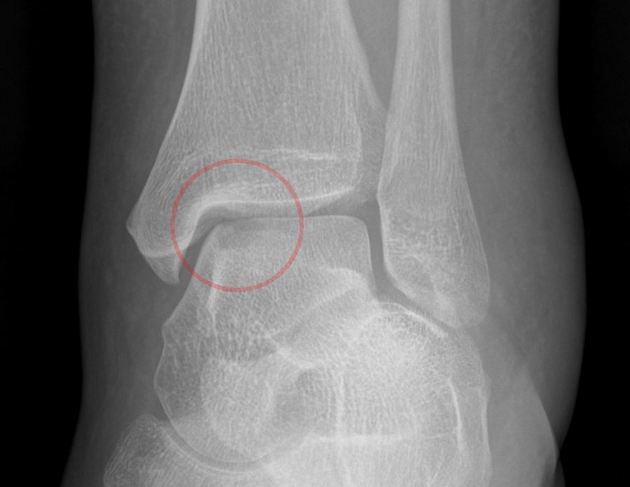
Red circle around the oval lucency on the oblique view, showing the fracture site.
Red circle around the fracture fragment on the lateral view (fragment arose from the talar dome). The fragment is located in the anterior joint space.
Case Discussion
Osteochondral defects/fractures can be nearly invisible on some radiographic projections of a joint, which is why "one view is no view"
These fractures also run a spectrum from a purely cartilaginous injury that can only be seen on MRI (type I) to this type of injury (type IV), in which the fragment is markedly displaced.


 Unable to process the form. Check for errors and try again.