
Presentation
Left-sided shoulder tenderness following a fall. Generalized shoulder pain on movement.
Patient Data

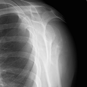
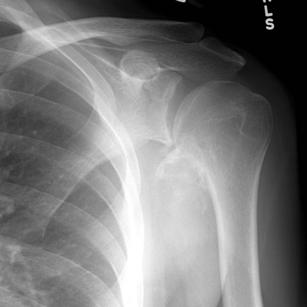
An expansile calcified / ossified mass arises from the proximal left humerus and projects medially. The base of the lesion appears continuous with the underlying bone. The medial and inferior parts of the lesion are ill-defined with a wide zone of transition.
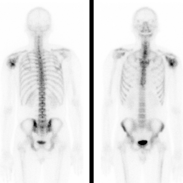
Focus of increased activity around the left shoulder in the region of the mass displayed on plain film. No other area of increased activity would suggest a solitary lesion.


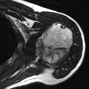
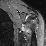
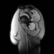
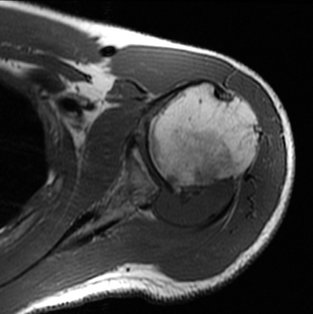
MRI confirms a lobulated soft-tissue mass arising from the posteromedial metaphysis of the left humerus.

Distribution of chondrosarcomas. Chondrosarcomas are widely distributed but are most frequently encountered in the femur, the proximal humerus and the pelvis, around the acetabulum. Layout and distribution: Frank Gaillard 2009, Line drawing of skeleton: Patrick Lynch 2006, Creative Common NC-SA-BY
The patient went on to have an en-bloc resection.
Left shoulder excision
Low grade (1/3) chondrosarcoma arising from a 7.5 cm osteochondroma of the posteromedial metaphysis of the humerus, with a maximal cartilage cap thickness of 2.4 cm (but also wrapping around much of the humeral head). Tumor extends into the glenohumeral joint space, but remains covered by reactive synovium. Negative for dedifferentiation.
Case Discussion
This case illustrates many of the features of a typical secondary chondrosarcoma arising from an osteochondroma. Recognizing the typical orientation and morphology of the underlying osteochondroma is key in making the diagnosis.


 Unable to process the form. Check for errors and try again.