
Patient Data


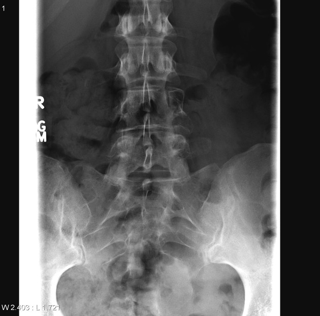
The L4 vertebral body appears somewhat lucent and the left pedicle is difficult to appreciate on frontal projection.
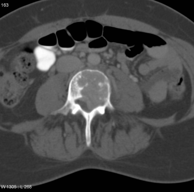
Lytic destructive lesion with matrix calcification and erosion of the posterior vertebral cortex and extending into the left pedicle noted in the L4 vertebra. There is extension into the spinal canal. No evidence of errosion of anterior and lateral margins of the vertebra.

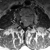
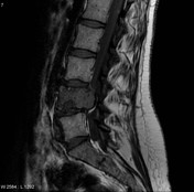

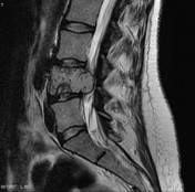
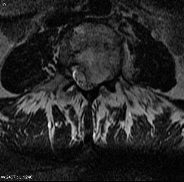
Heterogenous lesion that is predominantly hypointense on T1 and isointense on T2 with areas of calcification/coarse trabeculations seen as signal voids and heterogenous enhancement is seen in the body and left pedicle of L4, with erosion of the posterior cortex, but confined by posterior longitudinal ligament with no extension to soft tissue.
Case Discussion
The patient went on to have a complex resection of L4 vertebral body including left side posterior elements and associated soft tissues, adjacent intervertebral discs, the inferior body of L3 and superior body of L5.
Histology
Microscopic Description:
Sections show a clear cell neoplasm arranged as sheets and as lacunae within a cartilaginous matrix. Some woven bone is produced, osteoclasts are present and the lesion is more vascular than a conventional chondrosarcoma. Cells have abundant clear and faintly eosinophilic cytoplasm which is PAS positive in a strong, granular pattern. Nuclei are centrally-located and show low-grade atypia, with occasional mitoses identified. Immunostains demonstrate the lesional cells to be intensely positive for S100, but negative for keratins, EMA, synaptophysin and chromogranin.
Final diagnosis:
Clear cell chondrosarcoma (low-grade, grade 1/3)


 Unable to process the form. Check for errors and try again.