
Presentation
Diabetic foot ulcer. ? Osteomyelitis.
Patient Data

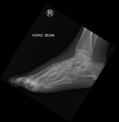
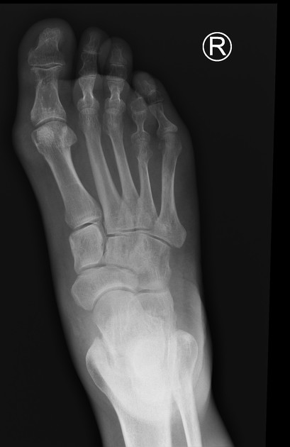
Erosion of the distal medial aspect of the first proximal phalanx with adjacent subtle increased density within the soft-tissue. Impression of increased density with soft-tissues medial to the first metatarsal head. The possibility of gout is raised as the cause for the proximal phalanx erosion.
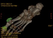

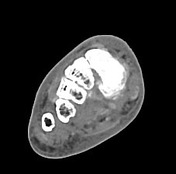
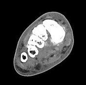
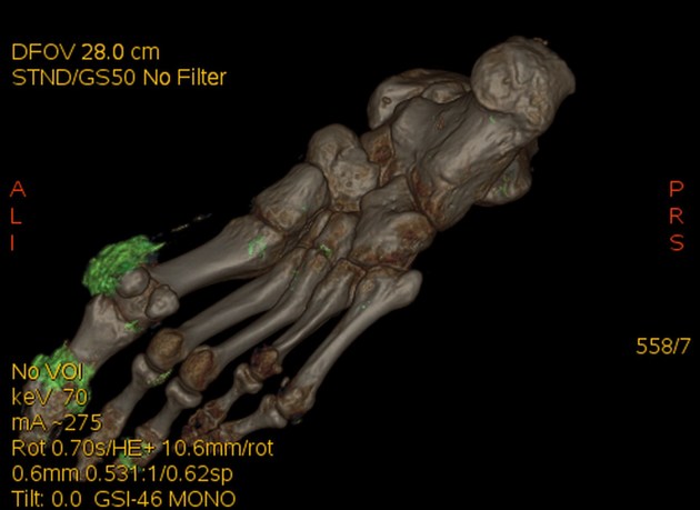
Generalized edema of the foot and ankle. Well-defined para articular erosion of the first proximal phalangeal head medially. Near circumferential to this articulation is a high attenuation crystal deposition corresponding to monosodium urate on dual energy CT. Further MSU deposition of the third DIP joint, second MTP joint, mid foot involving all of the TMT joints and the navicular-cuneiform articulation. There is also MSU deposition of the sinus tarsi and involving the lateral ankle ligament complex.
Case Discussion
A case of acute polyarticular gout confirmed with dual energy CT.


 Unable to process the form. Check for errors and try again.