
Presentation
History of recurrent forming renal stones with acute urine retention.
Patient Data

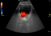

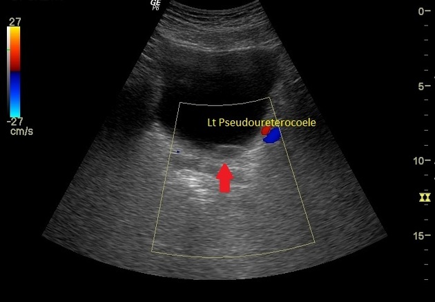
Distended urinary bladder, left-sided pseudo-ureterocele, intact ureteric jet bilaterally and normal size prostate.
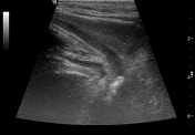
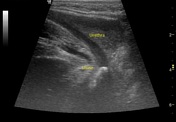
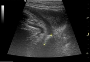
There is a well-defined hyperechoic structure with post acoustic shadowing located in the intraluminal part of the posterior urethra.

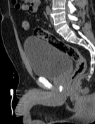
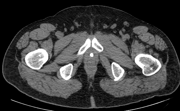
There is a hyperdense calcified structure in the posterior urethral course (posterior urethral stone) and distended urinary bladder (urinary retention).
Case Discussion
Acute urine retention in this age with a history of previous renal stones without a history of a chronic condition is highly suggestive of an impacted urethral calculus.
By using a linear probe in contact with the area between the scrotal sac and anus, this technique will visualize the posterior part of the urethra.
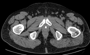
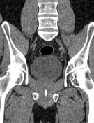
CT scan without contrast is the best modality to detect urolithiasis.


 Unable to process the form. Check for errors and try again.