
Presentation
Headache.
Patient Data
Age: 17 years
Gender: Female
From the case:
Empty sella


Download
Info
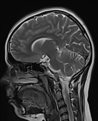
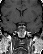
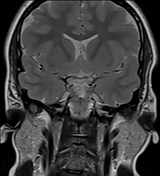
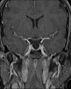
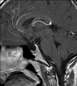
There is mild widening of the sella tursica , filled with CSF, with little enhanced pituitary tissue. The pituitary infundibulum is dipping in floor of the sella.
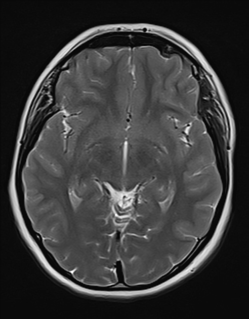
Also noted prominent optic nerve sheath more on left side is seen in axial T2W and sagital T2W images.
Case Discussion
The finding of an enlarged sella, filled with CSF, with the infundibulum traverse it is in keeping with an empty sella rather than a cystic lesion.
The prominent optic nerve sheath and relatively plump dural venous sinuses raise the possibility of underlying benign intracranial hypertension.


 Unable to process the form. Check for errors and try again.