
Presentation
Check position of Portacath.
Patient Data
Age: 45 years
Gender: Female
From the case:
Femoral Portacath
Download
Info
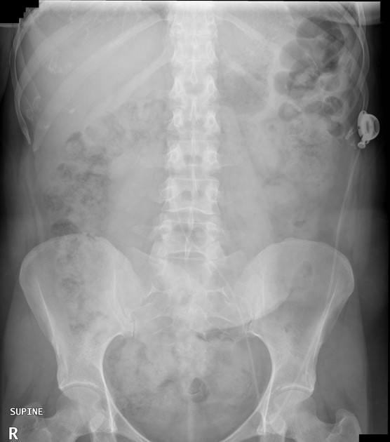
The left-sided abdominal Portacath tubing is intact and the tip is projected in the IVC at the level of the inferior endplate of L3. The bowel gas pattern is normal. No evidence of free gas.
Case Discussion
This patient had a history of right breast cancer requiring chemotherapy via a Portacath but her original catheter in her left chest became infected after one week and was removed. A catheter in the right side was contraindicated due to her surgical axillary clearance. She then had her treatment successfully completed via an Interventional Radiologist-inserted tunneled left abdominal Portacath with a common femoral vein puncture (under US-guidance).


 Unable to process the form. Check for errors and try again.