
Presentation
Pelvic pain and deformity.
Patient Data
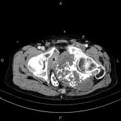

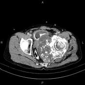
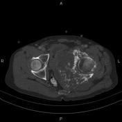

Large exophytic mass with a soft tissue component and chondroid calcification is seen on the left side of the pelvis, arising from the left ischiopubic and lower portion of iliac bones, measuring approximately 142 × 128 × 115 mm in diameter.
There is a marked mass effect on pelvic structures, with displacement of the rectum, urinary bladder, prostate gland, and seminal vesicles to the right.
The fat plane between the mass and urinary bladder and rectum is intact, but there is no clear fat plane between the mass and the lower aspect of the prostate gland. The proximal part of the left femur is intact.
Case Discussion
Pathology-proven case of chondrosarcoma in a 70-year-old male. On imaging, these tumors have rings and arcs chondroid matrix mineralization, with aggressive features such as lytic pattern, deep endosteal scalloping, and soft tissue extension.


 Unable to process the form. Check for errors and try again.