
Presentation
Mild dyspnea on exertion. Echocardiography showed septal hypertrophy.
Patient Data
Age: 35 years
Gender: Male
From the case:
Hypertrophic cardiomyopathy - septal and anterior pattern


Download
Info
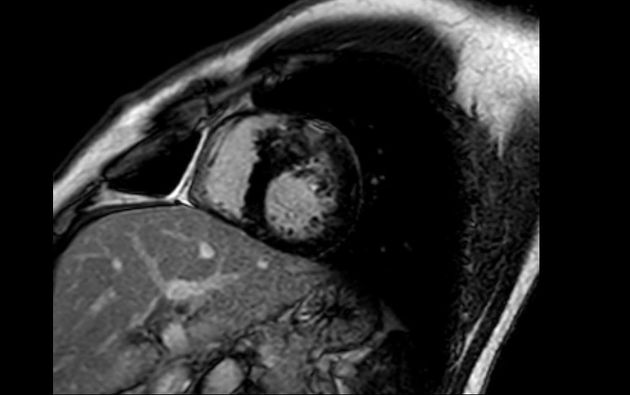
Sarcomeric hypertrophic cardiomyopathy with septal and anterior wall involvement is seen. There are also areas of patchy mid-wall enhancement indicating fibrosis. There is no left ventricular outflow tract obstruction.
Case Discussion
In this case there is hypertrophy of the septal and anterior wall but with sparing of the apex, which is less common than septal hypertrophy alone. The presence of fibrosis increases the risk of adverse cardiac outcomes.


 Unable to process the form. Check for errors and try again.