
Presentation
Abdominal pain.
Patient Data
Age: 35 years
Gender: Female
From the case:
Hepatic hydatid cyst


Download
Info
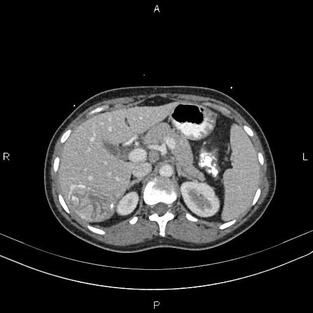
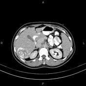
A 61 × 54 × 45 mm partially calcified cystic lesion is present in the right lobe of the liver contains multiple curvilinear hyper-attenuating internal structures in favor of inner layer membrane detachment.
Case Discussion
Features on the CT scan are compatible with hepatic hydatid infection.


 Unable to process the form. Check for errors and try again.