Presentation
Drowsiness, blurring of vision followed by coma. There was a suspicion of drug toxicity.
Patient Data
![](https://prod-images-static.radiopaedia.org/images/45956134/45af9b8604f82826760d86b97238c9_thumb.jpeg)
![This study is a stack](/packs/stack-YQKLCKBI.gif)
![](https://prod-images-static.radiopaedia.org/images/45956267/4298714d7dc3d5a6ae743ffa7db8dd_thumb.jpeg)
![This study is a stack](/packs/stack-YQKLCKBI.gif)
![](https://prod-images-static.radiopaedia.org/images/45956134/45af9b8604f82826760d86b97238c9_big_gallery.jpeg)
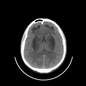
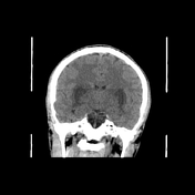
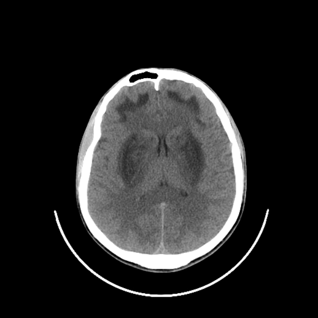
Bilateral symmetrical ill defined hypodensities involving the basal ganglia and subcortical white matter of both frontal lobes.
![](https://prod-images-static.radiopaedia.org/images/45956921/163705f6800cc25d4e4e1693134975_thumb.jpeg)
![This study is a stack](/packs/stack-YQKLCKBI.gif)
![](https://prod-images-static.radiopaedia.org/images/45957240/055bd59533cf8df022f6268accbeba_thumb.jpeg)
![This study is a stack](/packs/stack-YQKLCKBI.gif)
![](https://prod-images-static.radiopaedia.org/images/45956796/b38b0f377f7c2557f28565346b7e12_thumb.jpeg)
![This study is a stack](/packs/stack-YQKLCKBI.gif)
![](https://prod-images-static.radiopaedia.org/images/45957644/34a52b5d432268f33ced3c0df0b64a_thumb.jpeg)
![This study is a stack](/packs/stack-YQKLCKBI.gif)
![](https://prod-images-static.radiopaedia.org/images/45956654/cb5de83b7b9e5437f07a165b2a0e97_thumb.jpeg)
![This study is a stack](/packs/stack-YQKLCKBI.gif)
![](https://prod-images-static.radiopaedia.org/images/45956527/e6d5bf75648a632a48e2280fb844be_thumb.jpeg)
![This study is a stack](/packs/stack-YQKLCKBI.gif)
![](https://prod-images-static.radiopaedia.org/images/45957090/a95bffbb1ef151ad8b50b4ac7ea807_thumb.jpeg)
![This study is a stack](/packs/stack-YQKLCKBI.gif)
![](https://prod-images-static.radiopaedia.org/images/45957443/7c1336ad31760778d4a337e03584e9_thumb.jpeg)
![This study is a stack](/packs/stack-YQKLCKBI.gif)
![](https://prod-images-static.radiopaedia.org/images/45956921/163705f6800cc25d4e4e1693134975_big_gallery.jpeg)
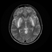
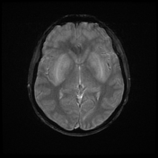
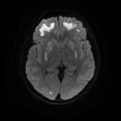
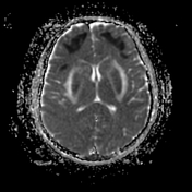
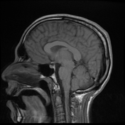
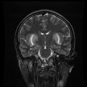
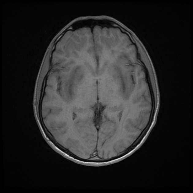
There was bilateral symmetrical signal alteration of the basal ganglia, mainly the putamen and caudate nuclei, and subcortical white matter of frontal regions displaying diffusion restriction, bright T2/FLAIR and low T1 signal. No hemorrhage was detected.
Left temporal and right parietal subcortical recent lacunar infarctions were noted.
Note complete opacification of the left maxillarly sinus, with secondary wall thickening, likely representing chronic sinusitis.
Case Discussion
After resuscitation of the patient from metabolic acidosis, he was found to have complete loss of vision of the right eye and poor vision of the left one. Ophthalmological examination revealed optic nerve atrophy.