
Presentation
Acute onset of bilateral lower limb weakness and parathesia. On examination, there was foot drop and sensory level.
Patient Data


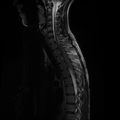

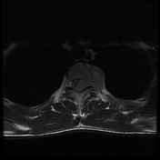
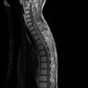

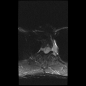
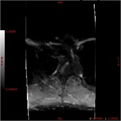
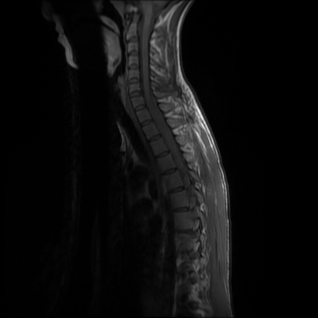
A longitudinal oriented intradural extramedullary soft tissue mass lesion is seen occupying the left side of the spinal canal opposite T1 down to T5 vertebrae, compressing the spinal cord to the right side and shows extra foraminal extension. The lesion elicits iso intense signal on T1 and hypointense signal on T2 with diffusion restriction on DWI and moderate homogeneous contrast enhancement. There is extra foraminal extension as well, the largest one through left T3-T4 foramen. Although the dorsal cord is compressed, but no significant intramedullary abnormal signal.
Case Discussion
The patient underwent surgical decompression and the lesion was found to be extra dural. Histopathology revealed Non-Hodgkin lymphoma.


 Unable to process the form. Check for errors and try again.