
Presentation
Low back pain and limping for 1 month following recent normal vaginal delivery
Patient Data
Age: 30 years
Gender: Female
From the case:
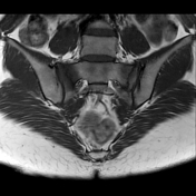
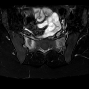
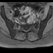
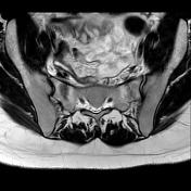
Sacral insufficiency fractures


Download
Info
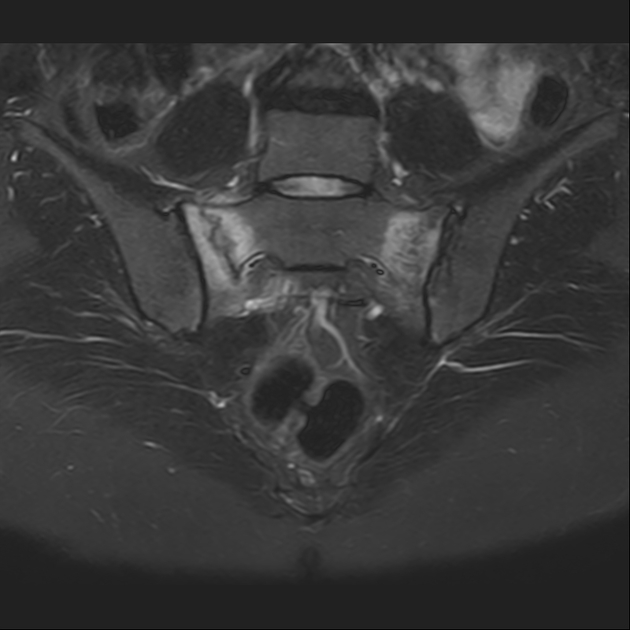
Linear irregular sacral fracture sclerotic lines parallel to the sacroiliac joints with surrounding bone marrow edema exhibiting bright signals on STIR & T2 fat sat images.
Mild bilateral subarticular bone marrow edema is seen at the sacroiliac joints, more at the iliac side on the right, denoting active sacroiliitis.
Download
Info

X-rays show no fracture lines.
Case Discussion
Sacral insufficiency fractures are a subtype of stress fractures occurring when normal stresses are applied to abnormal bone.
The patient had severe vitamin D deficiency, anemia, and recent normal vaginal delivery.
X-rays may be normal (as in this case).


 Unable to process the form. Check for errors and try again.