
Presentation
Patient treated for three weeks for a chest infection, presented with four days of repeated convulsions.
Patient Data
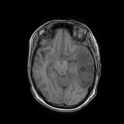

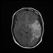
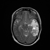
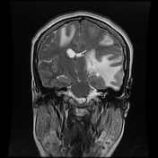
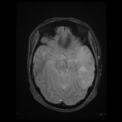
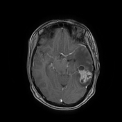
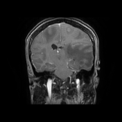
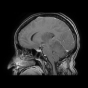
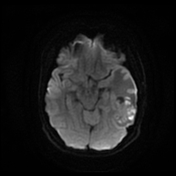


The MRI sequences demonstrate multiple intra-axial supratentorial lesions, one on, the right of frontal precentral location, and at least five on the left, the largest of temporal posterior location. These lesions are of low signal on T1WI, complex signal on T2WI with areas of low/high signal, surrounding vasogenic edema, and restricted diffusion on DWI. On postcontrast sequences, there is a peripheral ring enhancement of the small lesions and a cluster of ring enhancement of the largest lesion (granulomas). A mass effect is noted on the midline structures with subfalcine herniation.
The MRS (not shown) shows decreased NAA, and increased lipid/lactate.
Case Discussion
The MRI and MRS features in a patient treated for a chest infection (pulmonary tuberculosis in this case) are suggestive of intracranial tuberculous granulomas.
The patient's condition gradually improved with anti-tuberculous therapy.


 Unable to process the form. Check for errors and try again.