
Cerebral venous hemorrhagic infarct - cortical vein thrombosis
Presentation
Behavioral disorders with drowsiness.
Patient Data
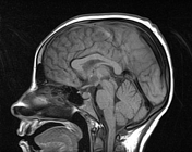

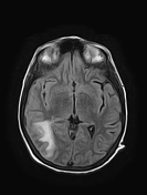
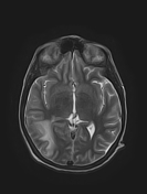
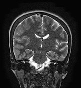
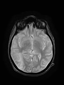
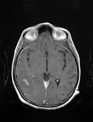
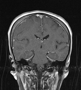
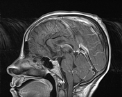
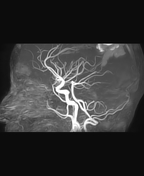
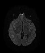
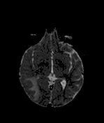
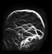
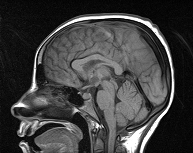
There is a right parietal hematoma mainly of cortical location, hyperintense on T1WI, and hypointense on T2WI/T2-GE with surrounding edema, extending to the posterior temporal lobe, and occipital lobe with mass effect on the adjacent occiptal horn. No enhancement seen on postcontrast sequences. Posterior to this hematoma, there is a dilated cortical vein of high signal on T1WI, and low signal on T*-GE (cortical thrombosis vein). The cerebral venous sinuses are patent on the MRV. No arteriovenous malformation or aneurysm is seen on the MRA. Note thickening with enhancement the pachymeninges, indicating most likely intracranial hypotension.
Case Discussion
Cerebral venous hemorrhagic infarct due to cortical vein thrombosis. Isolated cortical vein thrombosis without dural venous sinus involvement is reported to be extremely rare.


 Unable to process the form. Check for errors and try again.