Presentation
bilateral parotid enlargement treated as chronic parotitis.
Patient Data
![](https://prod-images-static.radiopaedia.org/images/48298197/78234a917c6ad434cd94743d752e23_thumb.jpeg)
![](https://prod-images-static.radiopaedia.org/images/48298188/ad546e109167f3eee91a3c1d77dc13_thumb.jpeg)
![](https://prod-images-static.radiopaedia.org/images/48298231/935713be22943e53e2d61326003599_thumb.jpeg)
![](https://prod-images-static.radiopaedia.org/images/48298216/9a0acce4b524dc123816b2b938c57f_thumb.jpeg)
![](https://prod-images-static.radiopaedia.org/images/48298239/46fb136895821f82f5a39c3db0fd6d_thumb.jpeg)
![](https://prod-images-static.radiopaedia.org/images/48298197/78234a917c6ad434cd94743d752e23_big_gallery.jpeg)
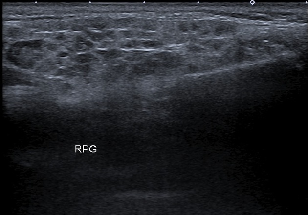
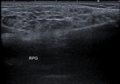
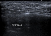
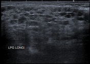
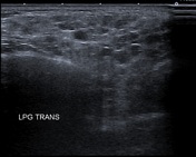
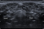
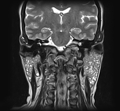
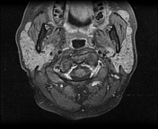
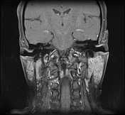
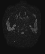
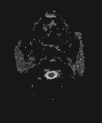
Both parotid and submandibular glands appear relatively enlarged with multicystic changes and reticular pattern.
![](https://prod-images-static.radiopaedia.org/images/48297059/b35d4def1235973747ad5978c2ea43_thumb.jpeg)
![This study is a stack](/packs/stack-YQKLCKBI.gif)
![](https://prod-images-static.radiopaedia.org/images/48297253/226db69d30db7b64517009fedd3eba_thumb.jpeg)
![This study is a stack](/packs/stack-YQKLCKBI.gif)
![](https://prod-images-static.radiopaedia.org/images/48296869/72058e74cf808dc860859d1d336476_thumb.jpeg)
![This study is a stack](/packs/stack-YQKLCKBI.gif)
![](https://prod-images-static.radiopaedia.org/images/48297443/b0b2d60252cbda8be778e4f5c3f621_thumb.jpeg)
![This study is a stack](/packs/stack-YQKLCKBI.gif)
![](https://prod-images-static.radiopaedia.org/images/48297635/b4c935dd8fc0454c6a04b7cb914872_thumb.jpeg)
![This study is a stack](/packs/stack-YQKLCKBI.gif)
![](https://prod-images-static.radiopaedia.org/images/48297829/357bdd037c9abdf81167b14a815f6d_thumb.jpeg)
![This study is a stack](/packs/stack-YQKLCKBI.gif)
![](https://prod-images-static.radiopaedia.org/images/48297949/b75c61c4cf4e1fc6d27b0386b80122_thumb.jpeg)
![This study is a stack](/packs/stack-YQKLCKBI.gif)
![](https://prod-images-static.radiopaedia.org/images/48297059/b35d4def1235973747ad5978c2ea43_big_gallery.jpeg)
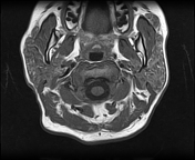
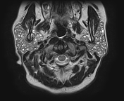
Both parotid glands are enlarged with innumerable cystic lesions disseminated in both parotid glands, of low signal onT1, hight signal on T2, giving a honeycomb appearance. On moderate heterogeneous enhancement is noted on postcontrast sequences.
Both submandibular glands show a similar but much less prominent involvement as compared to the parotid glands.
Case Discussion
Ultrasound and MRI features are characteristic of Sjögren syndrome.
Follow-up is mandatory in such case due to the high risk of developing a malignant lymphoma