
Presentation
Progressive headache, ataxia and recent blindness.
Patient Data
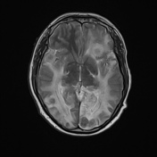

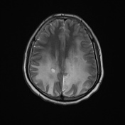
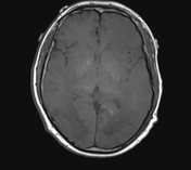
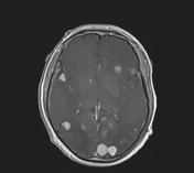
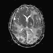
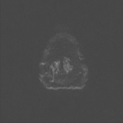
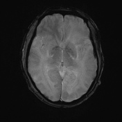
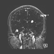
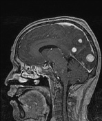

Multiple bilateral variable size supra tentorial intra-axial lesion in subcortical distribution showing low T1 signal, intermediated on T2 with homogenous enhancement associated with surrounding edema and white matter signal changes involving hypothalamic region, pons and dentate nuclei (high T2 and FLAIR) signal denoting neurodegenerative changes, no associated obstructive hydrocephalus, or skull bony lesions.
Case Discussion
This patient went on to have a biopsy. Histopathology revealed infiltration of the lesions by sheets of foamy histiocytes (xanthogranulomas) with plenty of multinucleated giant cells including Touton giant cells and chronic inflammatory cells rich in mature lymphocytes and few plasma cells with traversing short bands of spindle fibroblast-like cells. Findings were felt to be consistent with Langerhans cell histiocytosis.
Editorial note:
It is important to note that Touton giant cells are typically rare in Langerhans cell histiocytosis and thus the diagnosis in this instance should be questioned. Unfortunately, histological images and additional pathological investigations (e.g. electron microscopy) have not been provided. The diagnostic certainty has therefore been reduced to possible.
Langerhans cell histiocytosis is a systemic disorder involving the monocyte-macrophage system usually represent a systemic manifestation. CNS manifestation includes bony, intracranial hypothalamic-pituitary axis and pineal region, intracranial intra-axial lesions. The above case was initially diagnosed radiologically as CNS lymphoma or metastases.


 Unable to process the form. Check for errors and try again.