Presentation
Pre-op imaging before resection of a high-grade vulvar lesion.
Patient Data


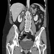
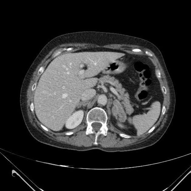
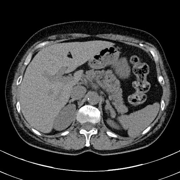
Bilateral thickening of adrenal glands, hypodense centrally with peripherally enhancing margins.
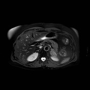
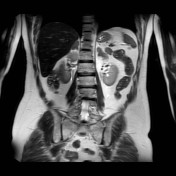
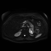
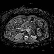
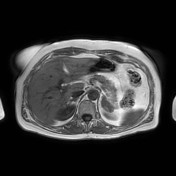
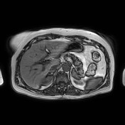
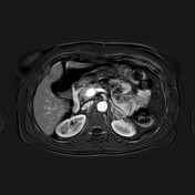


Slightly T2-hyperintense diffuse thickening of the adrenal glands with significant restricted diffusion and no signal drop between T1 in- and out-of-phase sequences. Subtraction images after contrast administration show only peripheral enhancement with central non-enhancement.
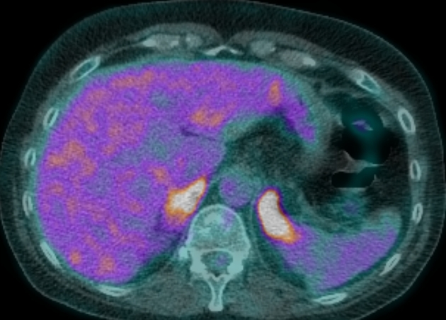
The adrenal thickening shows high FDG uptake on PET-CT.
Subsequent investigations, on the basis of ground-glass opacities on chest CT (not shown) revealed Histoplasma capsulatum on bronchoalveolar lavage.
Six months after treatment for histoplasmosis, the adrenal glands have returned to a normal appearance with resolution of the previously-noted hypodense thickening.
Case Discussion
Adrenal histoplasmosis can present with bilateral hypodense thickening of the adrenal glands on CT. Patients are at risk for subsequent adrenal insufficiency.


 Unable to process the form. Check for errors and try again.