
Presentation
Left posterior ankle pain.
Patient Data


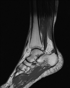
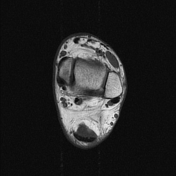

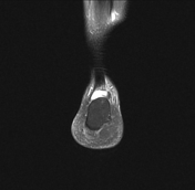

There is a fluid distended retrocalcaneal bursa with mild surrounding edema.
A linear hyperintense T2 signal is seen in the Achilles tendon 1.7 cm proximal to its insertion denoting tendinitis/tendinopathy. Small hyperintense T2 focus is seen at the insertion of Achilles tendon. There is also a thin hyperintense T2 signal around the distal aspect of the Achilles tendon suggesting peritendonitis.
No evidence of Haglund deformity. No thickening of Achilles tendon. The visualized bony structures showed normal bone marrow signal.
Case Discussion
Retrocalcaneal bursitis or insertional bursitis is inflammation of the retrocalcaneal bursa located anterior to the insertion of the Achilles tendon. It caused by infection, Achilles tendon injury, inflammatory arthropathies and calcaneal fractures.
It is usually associated with Achilles tendinitis and/or Haglund deformity.
It forms a part of Haglund syndrome (retrocalcaneal bursitis, Haglund deformity and Achilles tendinopathy).


 Unable to process the form. Check for errors and try again.