
Presentation
Proptosis. Abnormality seen on FDG-PET (not shown).
Patient Data
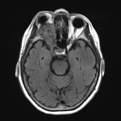

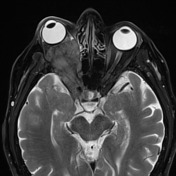
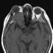

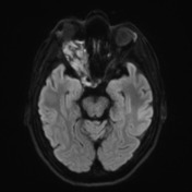
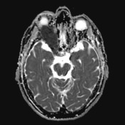
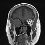
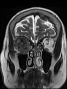
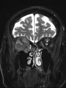
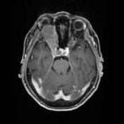
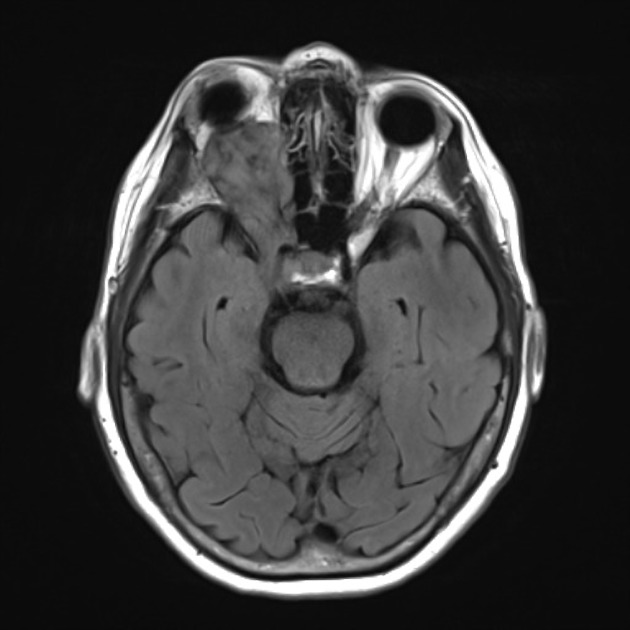
The region of marked FDG avidity centered in the right orbit corresponds to a lobulated, heterogeneously enhancing intra and extraconal mass which surrounds the right optic nerve to abut the posterior margin of the globe, without extension into the globe itself. The adjacent conus muscles are replaced or severely displaced by the mass, which progresses posteriorly through the orbital apex along the path of the right pre-chiasmatic optic nerve, almost entirely filling the right cavernous sinus, while preserving the right cavernous ICA flow void.
Tumor appears to extend beyond the roof of the right orbit suggesting extension beyond the periosteum intracranially.
Case Discussion
This individual had a known history of systemic carcinoid. They underwent enucleation.
Histology
Sections of globe show a well-differentiated neuroendocrine tumor within the soft tissue posterior to the eye. The tumor comprises infiltrative nests and sheets of cells with granular eosinophilic cytoplasm, ovoid nuclei and clumped chromatin. Mitoses are infrequent (2 per 42 HPF/10mm2) and there is no necrosis. There is evidence of some treatment effect with areas of hemorrhage, cholesterol clefts and foamy macrophages, however abundant residual tumor is present. Invasion of small vessels and small nerves is present.
There is infiltration of optic nerve sheath, however, invasion of the nerve or perineural space is not seen. Invasion of the globe is not seen and the sampled conjunctiva, sclera, iris and retina are unremarkable.
By immunohistochemistry, tumor cells are positive for synaptophysin, chromogranin and CD56. Ki67 proliferative index is 1-2%. 2-6.
Sections show woven trabecular bone and fibrous tissue infiltrated by well-differentiated neuroendocrine tumor with the same morphology
FINAL DIAGNOSIS: 1. Right eye: Well-differentiated neuroendocrine tumor (carcinoid)


 Unable to process the form. Check for errors and try again.