
Presentation
Metrorrhagia
Patient Data
Age: 50 years
Gender: Female
From the case:
Prolapsed endometrial polyp

Download
Info

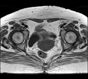
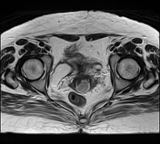
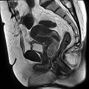
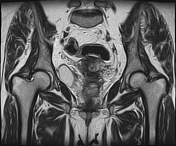
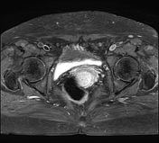
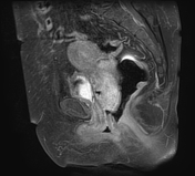
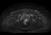
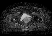
There is a large pedunculated intracavitary mass arising from the posterior uterine wall, filling the uterine cavity and prolapsing into the vagina through a distended endocervical canal. It is of intermediate signal intensity on T1WI, inhomogeneous high signal on T2WI surrounded by hyperintense fluid and endometrium with intense, and heterogeneous enhancement following IV contrast.
Case Discussion
MRI features are most consistent of a prolapsed endometrial polyp.
Additional contributor: Assia Kendri, radiographer, CIM Aurès, Batna Algeria


 Unable to process the form. Check for errors and try again.