
Presentation
Nuchal headache and visual disturbances.
Patient Data
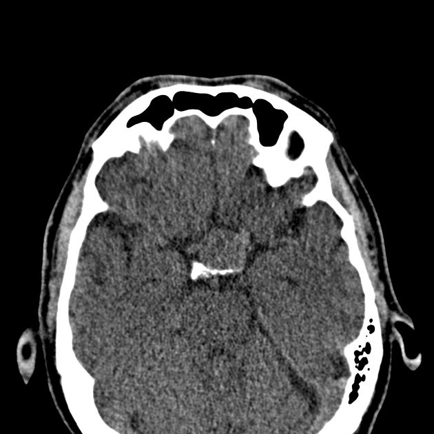
Noncontrast axial computed tomography (CT) shows the sella moderately enlarged, with an intrasellar well-defined mass, extending into the suprasellar cistern.


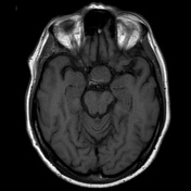
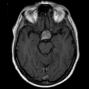


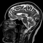
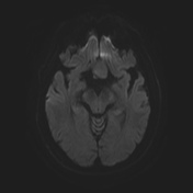
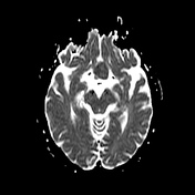
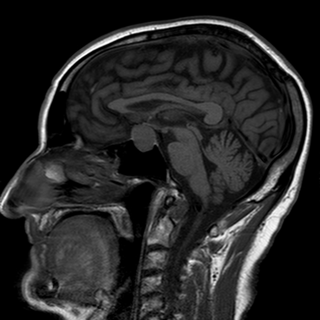
MRI demonstrates the enlarged pituitary fossa, filled by a solid enhancing tumor, extending into the suprasellar cistern, suggest pituitary adenoma. This tumor has overall dimensions of 1,7 x 2,1 x 2,2 cm. The mass causes severe chiasmal compression with superior displacement of more than 3 mm. On the left side, the lesion bulges into the cavernous sinuses, abutting the ipsilateral carotid artery, but not invading the carotid arteries on either side.
Case Discussion
Pituitary adenomas measuring more than 10 mm are macroadenomas. These tumors may cause visual impairment due to compression of the optic nerve, chiasm, or optic tract 1. Usually, pituitary macroadenomas higher than 2 cm in the vertical size 2 and displacement of the optic chiasm greater than 3 mm 1 produce vision impairment.
This case illustrates the typical appearance of a pituitary macroadenoma diagnosed based on clinical and imaging findings. This tumor was higher than 2,0 cm in vertical size, causing displacement of the optic chiasmal greater than 3 mm, and consequently vision disturbances on the patient.
Case courtesy
- Sterfferson Morais, MD - PGY-3, Radiology Resident, Department of Radiology
- Antonio Rodrigues de Aguiar Neto, MD - Radiologist, Department of Radiology
- Hospital da Restauração – Recife, PE – Brazil


 Unable to process the form. Check for errors and try again.