
Presentation
Severe headache, vomiting and unsteadiness.
Patient Data
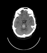

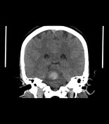
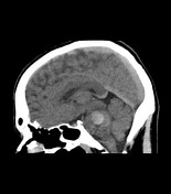

Right-sided pontine well defined hyperdense hemorrhagic lesion. It measures ~1.4 x 1.7 x 1.8 cm in maximum AP, TR & CC dimensions. It is surrounded by mild peri-focal edema. It exerts a mass effect in the form of compression and partially effaced right anterolateral aspect of the fourth ventricle. No intra-ventricular hemorrhage is present.

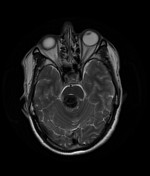
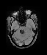
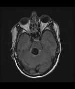
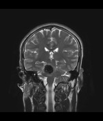
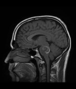
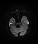
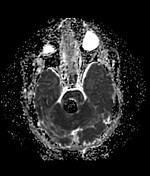
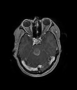
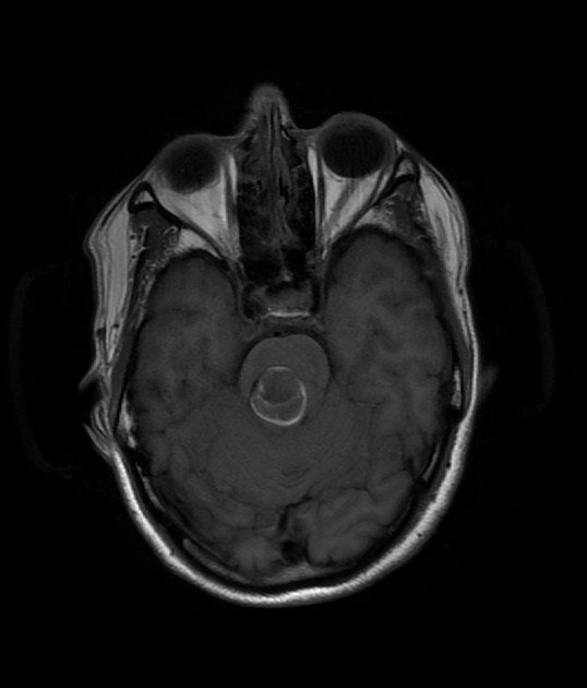
A predominantly acute bleed is seen at the right side of the pons is surrounded by mild perifocal edema. The GRE images confirmed the bleeding showing blooming. A small component of early subacute bleed is noted. Post-contrast images do not demonstrate underlying mass lesion with no significant post-contrast enhancement is seen. Underlying mass effect is noted with the effaced right anterolateral aspect of the fourth ventricle. No diffusion restriction of the pontine lesion is seen. No evidence of regional AVM or focal aneurysms. No hydrocephalus or CSF permeation is seen.
Case Discussion
Acute bleeding within a well-defined brain lesion is suggestive of cerebral cavernous venous malformation, commonly known as cavernous hemangioma or cavernoma, which is a common cerebral vascular malformation, usually with characteristic appearances on MRI. Cavernous malformations of the brainstem are uncommon, but the natural history appears to be worse than those in other locations. Retrospective annual hemorrhage rates appear to be around 5%.


 Unable to process the form. Check for errors and try again.