Presentation
Headache
Patient Data


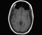
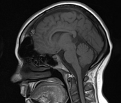
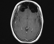

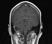

Bilateral symmetrical dilatation of both frontal sinuses with normal aeration. Normal size of the otherwise paranasal sinuses.
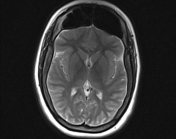
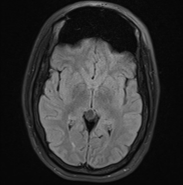
A pineal cyst is noted with fluid content similar to CSF in all pulse sequences.
Case Discussion
Pneumosinus dilatans refers to an isolated enlarged aerated paranasal sinus (hyperpneumatisation) with intact bony walls and bony septae. It is most commonly seen in frontal sinuses. It is a normal variant and usually asymptomatic with incidental discovery.
A pineal cyst is also an incidental finding. Here it shows typical appearance with fluid content similar to CSF. It can show variable T1 signal according to its content. Cystic pineocytoma may have similar imaging appearance and follow up is recommended for cysts larger than 10 mm.


 Unable to process the form. Check for errors and try again.