
Presentation
Fever for 4-5 days. Altered sensorium and vomiting.
Patient Data
Age: 20 years
Gender: Female
From the case:
Japanese encephalitis


Download
Info
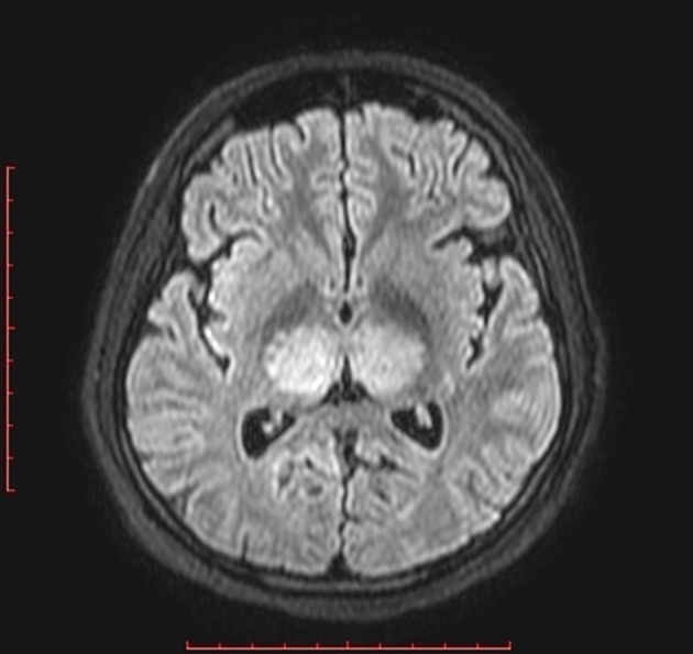
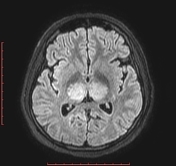
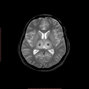
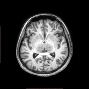
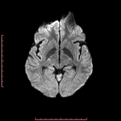
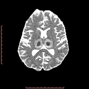
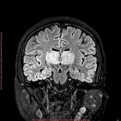
Hyperintense signal on T2 and FLAIR images involving bilateral thalami, caudate nuclei, pons, midbrain, left occipital lobe and bilateral cerebellar hemispheres. Patchy focal areas of blooming are seen within the thalami and pons, suggestive of hemorrhage. Basilar artery flow void is maintained on T2 images. Venography showed no thrombosis of the internal cerebral vein or straight sinus.
Case Discussion
Japanese encephalitis still remains a major health problem in India and Southeast Asia. Clinical history with typical MRI features showing bilateral thalamic involvement are pivotal in diagnosis.


 Unable to process the form. Check for errors and try again.