
Presentation
Neck swelling.
Patient Data
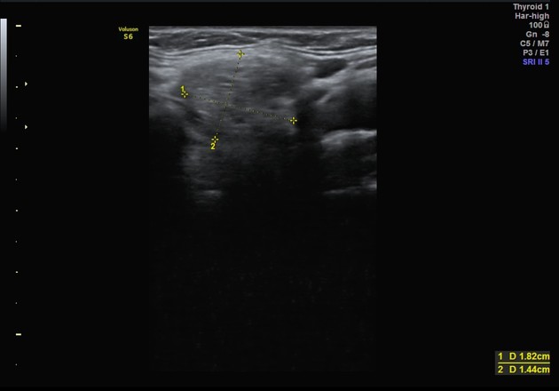
Enlarged thyroid gland with heterogeneous nodular echo pattern and normal color flow on Doppler. The small rounded structure is seen anteriorly to the related to strap muscle at the level of the hyoid bone. It may represent ectopic thyroid tissue.


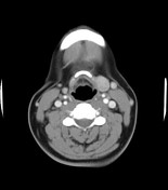
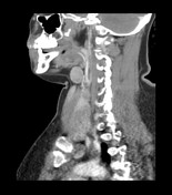
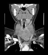
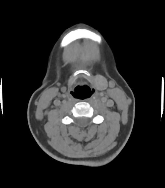
The thyroid gland is diffusely enlarged with a nodular heterogeneous CT attenuation and scattered calcific foci. At the level of the hyoid bone and to the left, closely related to the strap muscles, is a well-defined rounded structure with hyperdense, slightly heterogeneous CT attenuation similar to the thyroid. It measures 1.6 x 1.9 x 2.2 cm. The appearance is most suggestive of an ectopic thyroid gland. Bilateral small multilevel cervical lymph nodes.
Case Discussion
The ultrasound and CT findings are suggestive of ectopic thyroid tissue. Further assessment would be a thyroid scan to confirm our radiologic diagnosis.


 Unable to process the form. Check for errors and try again.