
Presentation
Diffuse abdominal pain and increased abdominal volume. On physical examination, a palpable mass extending from the right hypochondrium to the ipsilateral iliac fossa.
Patient Data
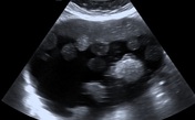

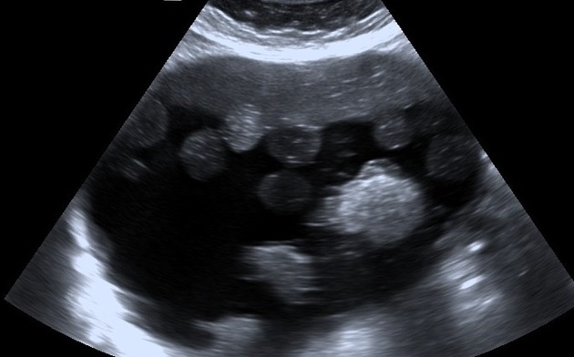
Voluminous formation containing numerous echogenic balls floating in an anechoic liquid.
In the posterior region, there are echogenic structures with posterior acoustic shadow probably due to calcifications, hair strands, or foci of fat.

Macroscopic appearance after surgery and liquid drainage of the ball; bulky saccular formation containing fat balls and hairballs.
Case Discussion
Ovarian teratoma is one of the most frequent benign ovarian tumors of reproductive age in women. When mature and cystic, it may also be called a dermoid cyst.
The appearance of cysts containing multiple internal floating balls is a very rare presentation of the dermoid cyst, but is considered pathognomonic for this entity and was first described in 1991 1,2. These internal floating balls may be single or multiple, measuring around 5 to 40 mm, composed of keratin, fibrin, hemosiderin, hair, and fat in different proportions and their mechanism of formation has not been well defined yet.
This imaging aspect is not unique to ovarian teratomas, it can be seen in the rectouterine pouch, mediastinum, and skull teratomas. They are usually asymptomatic and slow-growing but may manifest with discomfort and abdominal bloating, sometimes progressing to rupture, malignant degeneration or causing compressive symptoms due to the expanding size 3-6. Torsion cases are also reported, but mainly in larger than average dermoid cyst (average diameter of 11 cm) 7. Although ultrasound findings have been reported to be sufficient for diagnosis. Complementary examinations such as computed tomography and magnetic resonance imaging are often necessary to define the origin of the lesion.


 Unable to process the form. Check for errors and try again.