
Presentation
Past history of trauma, difficulty in breathing
Patient Data
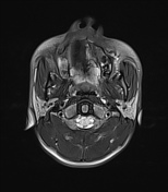

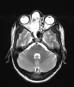
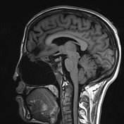


Large defect in the cribriform plate of ethmoid on right side measuring approx. 7.5 mm on transverse and 10 mm in anteroposterior dimension with herniation of cerebral parenchyma and meninges through it.
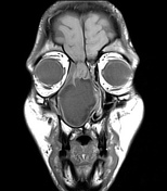
Associated large cystic lesion, measuring approx. 67 x 43 x 50 mm (AP x TD x CC) showing CSF signal intensity is seen occupying nasal cavity, causing significant expansion, scalloping of nasal bone, with deviation towards left side and compression of left nasal cavity. It is causing scalloping of medial wall of right maxillary sinus with mild compression of sinus.
The lesion is abutting palate inferiorly and obliterating nasopharyngeal airway posteriorly.
Gliotic area is seen involving right basifrontal lobe causing mild ex-vacuo prominence of frontal horn of right lateral ventricle.
Case Discussion
Findings are consistent with large meningoencephalocele.


 Unable to process the form. Check for errors and try again.