
Presentation
Persistent superomedial knee pain.
Patient Data
Age: 35 years
Gender: Male
From the case:
Medial patellar plica syndrome


Download
Info

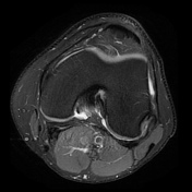
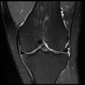
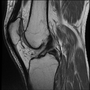
Thickened medial patellar synovial plica interposed between the medial patellar facet and medial femoral trochlea. Adjacent multifocal near full-thickness chondral fissuring of the medial patellar facet. Articular cartilage of the patellofemoral compartment is otherwise preserved.
Case Discussion
Medial patellar plica syndrome is an uncommon cause of anterior knee pain. Here, the MRI findings correlate strongly with the clinical presentation.


 Unable to process the form. Check for errors and try again.