
Presentation
Recent headaches.
Patient Data
Age: 60 years
Gender: Female
From the case:
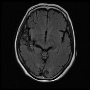
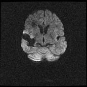
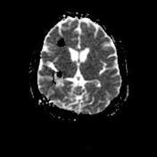
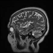
Multiple cerebral cavernomas


Download
Info
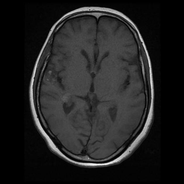
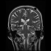
Multiple scattered T2 centrally hyperintense lesions with thin T2 hypointense rim in the right cerebral hemisphere, left occipital lobe and right cerebellar hemisphere with no perilesional gliosis and edema.
The majority of the lesions also show focal intralesional T1 hyperintense signal in keeping with a more recent bleeding cavernomas.
No mass effect or midline shift.
Case Discussion
Cavernous malformations are usually isolated. Where multiple cavernous malformations are present, familial multiple cavernous malformation syndrome must be considered. This condition typically presents with seizures. The inheritance pattern is autosomal dominant, with variable penetrance.


 Unable to process the form. Check for errors and try again.