
Presentation
Right scrotal swelling
Patient Data
Age: 55 years
Gender: Male

Download
Info
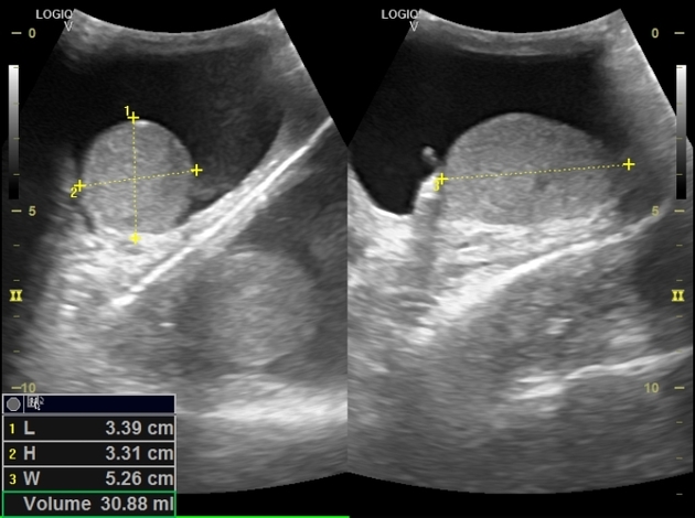
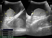
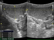
Moderate right hydrocele with clear fluid.
Few small right epididymal cysts averaging 5 mm.
Case Discussion
The first stack (scanning through right lateral scrotal wall) shows suspected bilateral scrotal hydrocele which confirmed not to be true on scanning through the anterior approach (second stack). The scrotal septum, in our case, acted as a highly reflective surface that formed the false image. To avoid this, scan in different angles and positions to change the angle of insonation.


 Unable to process the form. Check for errors and try again.