
Presentation
Known case of NF1
Patient Data


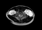
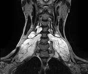
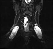
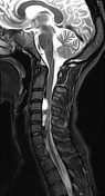
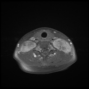
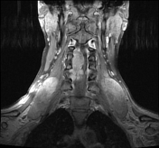
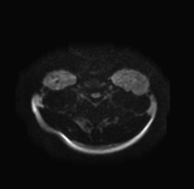
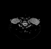
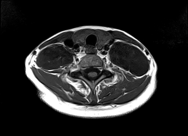
The MRI sequences demonstrate bilateral cervical masses in the distribution of the nerve roots, extending from C3-C4 up to C6-C7. The largest masses are at C5-C6. with compression of the adjacent segment of the spinal cord from C3-C4 to C5-C6 level. They are of low signal on T1WI, high signal on T2WI with moderate and heterogeneous enhancement on postcontrast sequences. Note vertebral scalloping mainly of C4, C5, and C6.
Case Discussion
MRI features of bilateral plexiform neurofibromas in a patient with neurofibromatosis type 1
Plexiform neurofibromas are considered as benign tumors of peripheral nerves (WHO grade I), arising from a proliferation of all neural elements. They are seen in approximately 30% of patients with NF1 and considered pathognomonic of this disease.


 Unable to process the form. Check for errors and try again.