
Presentation
A twisting injury a few weeks ago. Clinically anterior instability, extension deficit.
Patient Data
Age: 45 years
Gender: Male
From the case:
Radial meniscal tear and ACL injury


Download
Info
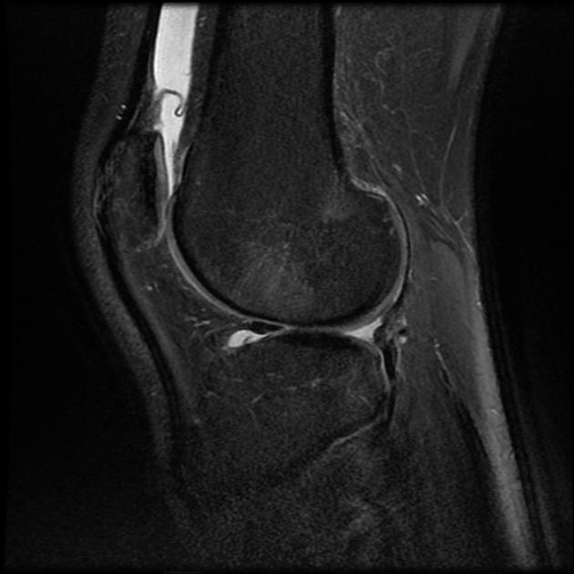
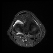
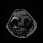
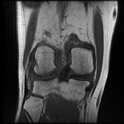
There's a high-grade partial ACL tear, with torn fibers rolled up anteriorly to the Hoffa fat pad and concomitant bone bruises at locations typical for a pivot-shift injury. A complete radial tear of the posterior horn of the lateral meniscus is also visible, with separation of the torn edges and meniscal body extrusion.
Case Discussion
When a large radial meniscal tear transects the meniscus completely, it gives the appearance of a 'ghost meniscus' on sagittal MR images, with no visible meniscus on slices that course through the fluid gap.


 Unable to process the form. Check for errors and try again.