
Presentation
Road traffic accident 1 month ago.
Patient Data
Age: 25 years
Gender: Male
From the case:
Hoffa fracture of the lateral femoral condyle


Download
Info
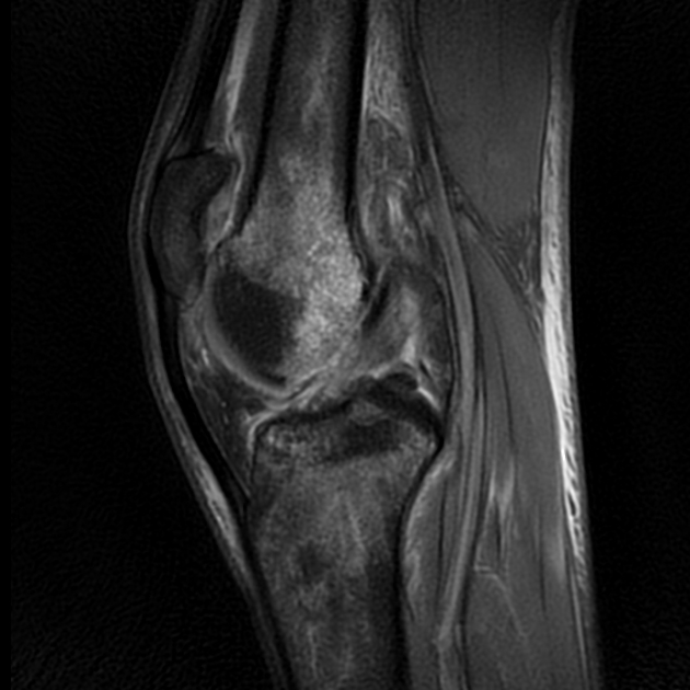
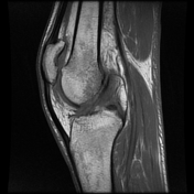
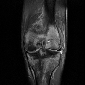
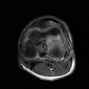
The MRI sequences demonstrate a slightly displaced posterior, intra-articular fracture of the lateral femoral condyle with extensive bone marrow edema and joint effusion. Note an associated fibular head fracture.
Menisci, cruciate and collateral ligaments appear intact.
Case Discussion
MRI features of Hoffa fracture of the lateral femoral condyle with associated fibular head fracture.


 Unable to process the form. Check for errors and try again.