
Presentation
Pelvic pain and tenderness
Patient Data


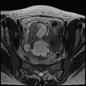
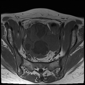
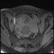
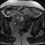
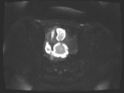
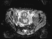

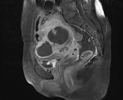
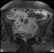
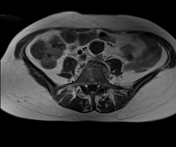
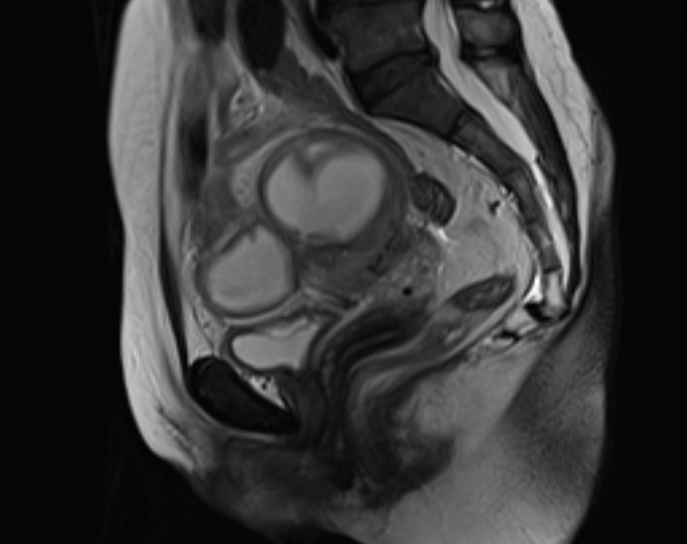
A tubular/cystic right adnexal lesion is noted, measuring 8.5 x 10.5 cm, showing thick enhancing walls and non-enhancing fluid contents showing marked Diffusion restriction (suggesting purulent nature) with no solid components. Marked pelvic inflammatory fat stranding.
Dilated right ureter (9 mm) mostly sequel to this abscess.
IUCD in place.
Case Discussion
This case demonstrates features of pelvic inflammatory disease (PID) with right tubo-ovarian abscess with pyosalpinx and pelvic peritonitis. The ovaries couldn't be distinguished separately from the fallopian tubes.
A tubo-ovarian abscess is a late complication of pelvic inflammatory disease (PID). It is important to differentiate it from other causes of pelvic abscess like appendiceal abscess and diverticular abscess. MRI is the best choice if clinically suspecting a pelvic inflammatory mass.


 Unable to process the form. Check for errors and try again.