
Presentation
Acute visual loss and headache.
Patient Data


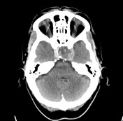
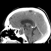
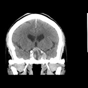
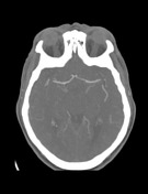
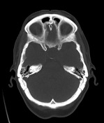
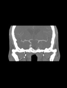
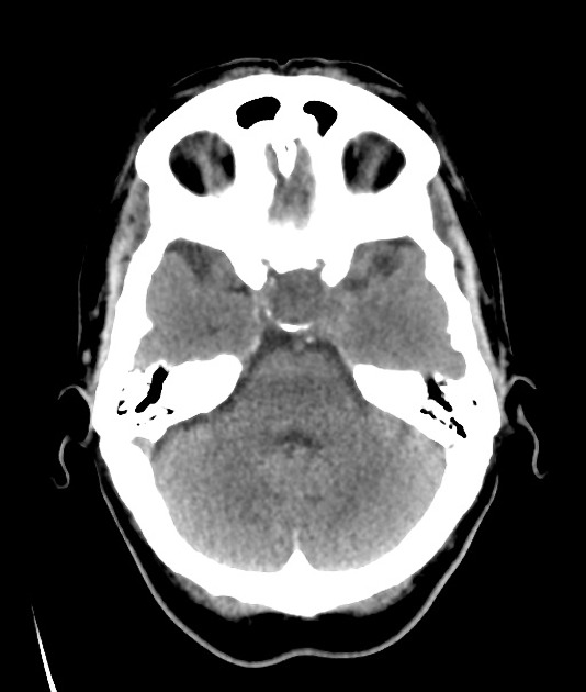
The pituitary fossa is expanded and remodeled by a sizable mass that has mixed density including some central components that are low and moderately highly attenuating.

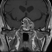

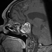
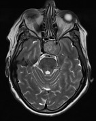
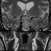
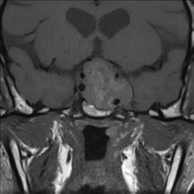
A pituitary region mass is present that invades the left cavernous sinus without arterial stenosis. Suprasellar extension is present that compresses the optic chiasm. The mass is heterogeneous enhancing with central non-enhancement which has intrinsic high T1 signal and areas of low and high T2 signal, in keeping with blood.
Case Discussion
The patient went on to have surgery.
Histology
The sections show moderately cellular pituitary adenoma comprising sheets and nests, surrounded by vascularized stroma. The tumor cells have mildly enlarged round nuclei, finely granular chromatin and moderate amounts of eosinophilic cytoplasm. Elsewhere, there is evidence of apoplexy with tumor necrosis and hemorrhage. Granulation tissue with scattered chronic inflammatory cells is present. There is a small amount of normal anterior pituitary gland tissue present.
Scattered tumor cells are weakly positive for growth hormone. Entrapped prolactin positive cells are noted. Immunostains for the other pituitary hormones are negative. The Ki-67 index is difficult to determine as there are scattered positively stained inflammatory cells.
FINAL DIAGNOSIS: Somatotroph adenoma with evidence of apoplexy.


 Unable to process the form. Check for errors and try again.