
Presentation
Headache
Patient Data


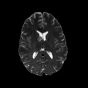
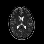
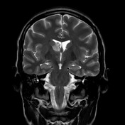
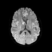
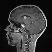
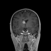
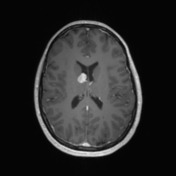
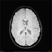
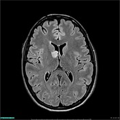
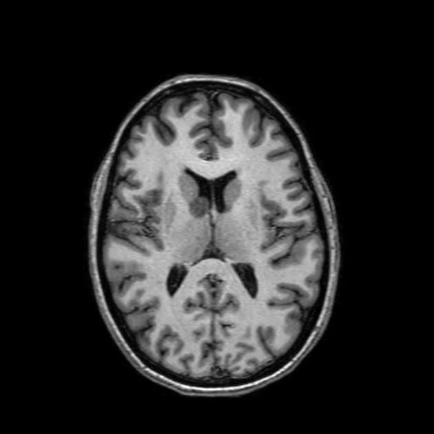
A lobulated vividly enhancing mass is located near the right foramen of Munro. It is an isolated abnormality. No subependymal nodules or parenchymal T2 signal abnormalities.
Case Discussion
The patient went on to have a resection.
Histology
Sections show pieces of tumor of relatively low cellularity. The tumor cells are dispersed within a fibrillary background and form vague fascicles and sheets. The majority of the cells show marked enlargement and prominent pleomorphism. They have large oval-to-irregularly shaped eccentric hyperchromatic nuclei with large amounts of homogeneous eosinophilic cytoplasm. There are scattered very large multinucleated cell is also seen. There are interspersed small cells that have a spindled or epithelioid morphology. There are small lymphocytes and mast cells scattered throughout the tumor. There are no mitoses identified and no necrosis present. There are scattered parenchymal calcifications.
IMMUNOHISTOCHEMISTRY:
- GFAP: variably GFAP positive, with strong expression in a subset of tumor cells
- IDH1 R132H: negative (non-mutated)
- ATRX: positive (non-mutated)
- p53: patchy weak
- p16: negative
- Topoisomerase: less than 1%
- S100: diffusely positive in tumor cells
- c-kit: stains intratumoral mast cells
- Mast cell tryptase: stains intratumoral mast cells
- NFP: stains a few cellular processes and the occasional tumor cell cytoplasm
- NeuN: negative
FINAL DIAGNOSIS: subependymal giant cell astrocytoma - WHO grade I.
Discussion
This case is particularly interesting as the patient had no features of tuberous sclerosis either within the brain or systemically.
Sporadic subependymal giant cell tumors are rare but reported.


 Unable to process the form. Check for errors and try again.