
Presentation
Hematology patient. Headache. Missed anticoagulants.
Patient Data
Age: 20 years
Gender: Female
From the case:
Dural sinus thrombosis - empty delta sign

Download
Info
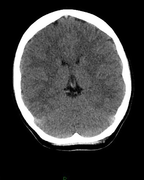
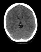
Hyperdense posterior superior sagittal sinus on the non contrast images.
The patient was immediately recalled from accident and emergency for a CT of the venous sinuses.
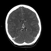
Large filling defect in the posterior superior sagittal sinus, corresponding with the hyperdense area on the non contrast images. The filling defect extends into the right transverse sinus.
Filling defect in the right internal jugular vein and sigmoid sinus.
No parenchymal hemorrhage or venous infarct.
Case Discussion
The delta (non-contrast study) and empty delta (contrast study) signs are exquisitely demonstrated on this CT study.


 Unable to process the form. Check for errors and try again.