
Presentation
Presented with clinical picture of right orbital cellulitis.
Patient Data
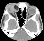
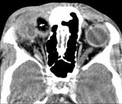


CT scan shows cystic lesion in the extraconal superomedial aspect of the right orbit. It contains fat and fluid. Perilesional inflammatory changes are noted which extends into the upper lid. The cyst wall appears thick, enhancing with focal point of interruption (arrow).
Case Discussion
This case illustrates an orbital dermoid cyst complicated by rupture and acute inflammation.
Orbital dermoid usually presents as a painless subcutaneous mass.
If the cyst ruptures, either spontaneously or with trauma, a pronounced inflammatory response will occur that may mimic orbital cellulitis or inflammatory rhabdomyosarcoma. The inflammation may be suppressed by corticosteroids, but excision is required to prevent recurrence.


 Unable to process the form. Check for errors and try again.