
Presentation
Longstanding headache and dizziness as well as paresis.
Patient Data


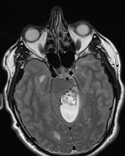
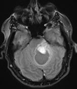
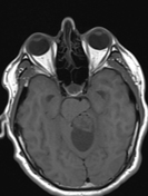
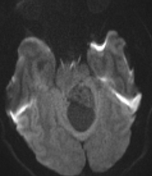
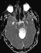
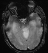
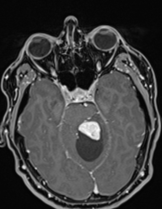
A large cystic mass lesion is seen in the posterior cranial fossa mainly vermis and left cerebellar hemisphere containing enhancing soft tissue solid component with two foci of necrosis as well as few tubular flow void within the solid component.
The lesion surrounded by edema extending to the brainstem also causing mass effect on the brainstem and producing tonsillar herniation, ascending transtentorial herniation and active hydrocephalus.
Case Discussion
Hemangioblastomas are uncommon tumors of the central nervous system, corresponding to 1–2.5% of all intracranial tumors. They can present sporadically or in patients with von Hippel–Lindau disease and are most often located in the cerebellum, brainstem, and spinal cord.1


 Unable to process the form. Check for errors and try again.