
Presentation
The patient presented for a CT scan because the referring clinician suspected left renal agenesis.
Patient Data
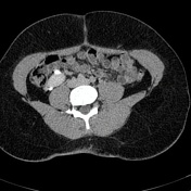

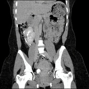
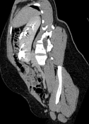

The left kidney is not visualized in its normal anatomical position but is instead crossed over to lie ipsilateral to the right kidney, to which it is fused at its inferior pole. The homogenous density across the two fused renal poles suggests the presence of a parenchymal band joining the two kidneys.
Both kidneys are malrotated, and each has its own renal pelvis.
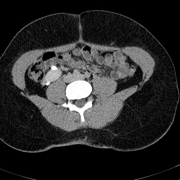
The ureter of the left ectopic kidney crosses the midline anterior to the right common iliac vessels and enters the bladder on the opposite side. The left vesicoureteric junction appears to be ectopically located, more inferiorly than normal.
The right ureter has a more normal course, lying adjacent to the right psoas muscle. The right VUJ also has a more normal position compared to the left.
There is no calculus nor mass lesion seen within the urinary system and no evidence of hydronephrosis.
The urinary bladder is normal in appearance.
Case Discussion
Crossed fused renal ectopia is the second most frequently observed type of renal fusion anomaly following the horseshoe kidney, where one kidney crosses the midline to lie on the same side as the other kidney, as well as fuse with it. The ureter of the crossed kidney will however still insert onto the urinary bladder on the opposite (correct) side.
Therefore, in this case left-to-right renal ectopy is observed, which is thought to be more common (3 times more) than the other way around.
Several subtypes or variations of the crossing and fusion exist including inferior crossed fused ectopia (as in this case), superior crossed fused ectopia, sigmoid, lump, disc, and L-shaped kidneys.
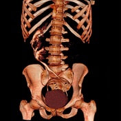
In any type of crossed fused ectopic kidney, the vascular anatomy should be closely studied as it is often grossly aberrant, while the crossed ectopic kidney also often demonstrates decreased function.


 Unable to process the form. Check for errors and try again.