
Presentation
Raised inflammatory markers with no clear cause
Patient Data
Age: 65 years
Gender: Male


Download
Info
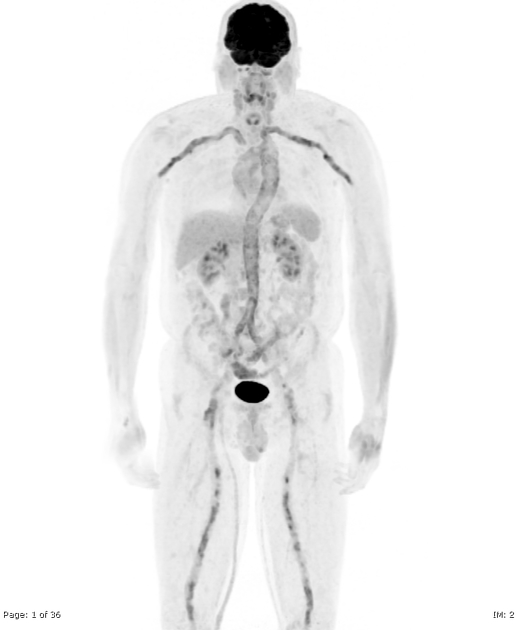
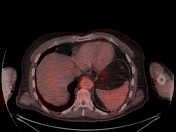
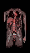
Moderate to marked increased tracer uptake is seen throughout the aorta, iliac, femoral, subclavian and axillary arteries reflecting an active large vessel vasculitis.
Case Discussion
This case demonstrates the typical distribution of active large vessel vasculitis on FDG PET-CT.
EANM/SNMMI guidelines for vasculitis in FDG PET-CT:
•Grade 0: No vascular uptake (≤ mediastinum)
•Grade 1: Vascular uptake < liver uptake
•Grade 2: Vascular uptake = liver uptake, may be PET-positive
•Grade 3: Vascular uptake > liver uptake, considered PET-positive


 Unable to process the form. Check for errors and try again.