
Presentation
Left breast mass. Two months post partum.
Patient Data
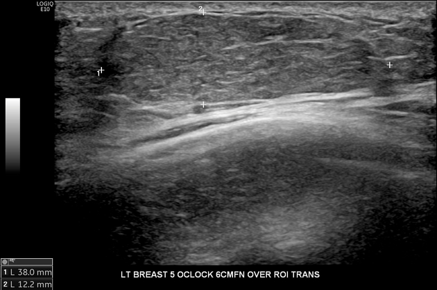
The palpable mass correlates to a 38 x 27 x 12 mm mass with smooth margins and similar echogenicity to the adjacent breast parenchyma. No vascularity.
Case Discussion
This patient self-detected the lump in her third trimester of pregnancy, and since breastfeeding, the lump got larger. The differential diagnosis was fibroadenoma, hamartoma and lactating adenoma.
The patient underwent an ultrasound-guided core biopsy.
Histopathology
MACROSCOPIC DESCRIPTION: Four tan needle cores 3-14 mm.
MICROSCOPIC DESCRIPTION: The sections show core biopsies of fibrofatty breast tissue containing benign breast lobules demonstrating lactational change. There is no evidence of dysplasia or malignancy.
CONCLUSION:
Breast, left, 5 o'clock core biopsy: Lactating adenoma (nodular lactational hyperplasia).


 Unable to process the form. Check for errors and try again.