
Presentation
Acute onset bilateral upper and lower limb weakness.
Patient Data


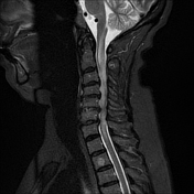
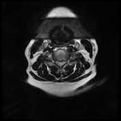
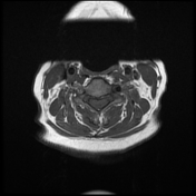
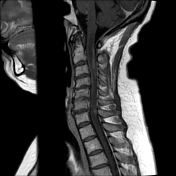
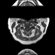

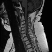
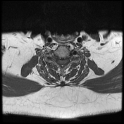
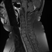
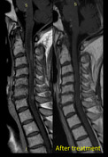
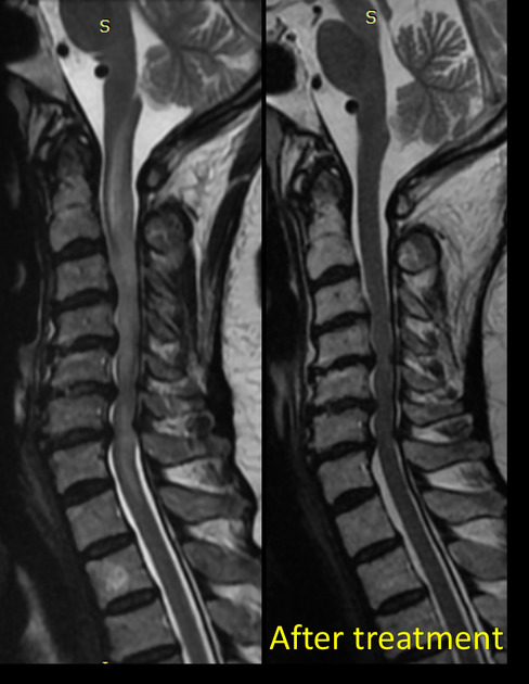
The cervical cord is expanded over a long segment between C2-T1 level with diffuse symmetrical high signal change on T2/STIR, involving both sides of the cord. Associated multiple intramedullary patchy areas of abnormal enhancement on postcontrat sequences.
Cervical spondylodegenrative changes.
C3-4 central posterior disc protrusion.
C4-5 posterior and left posterolateral disc protrusion.
C5-6 and C6-7 diffuse posterior disc bulges with central protrusions.
Bilateral C4-5, C5-6 & C6-7 degenerative neurocentral arthropathy.
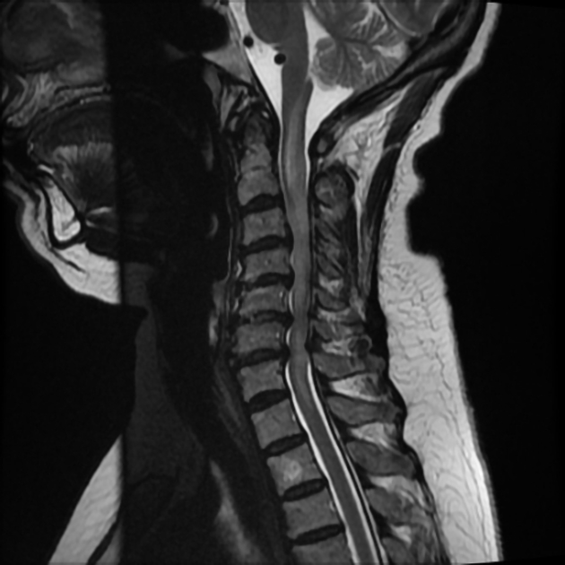
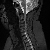
MRI of the dorsal spine showed normal appearance of the dorsal spinal cord.
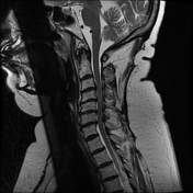
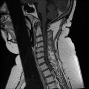

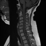
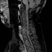
Follow-up after corticosteroid treatment showed complete resolution of the cervical cord swelling and patchy contrast enhancement.
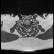
Stationary course regarding the cervical spondylosis and disc lesions.

Follow-up after corticosteroid treatment showed complete resolution of the cervical cord swelling and patchy contrast enhancement.
Case Discussion
Clinical and first MRI were both favoring the diagnosis of transverse myelitis, after which the patient received corticosteroid treatment. The patient showed clinical improvement after treatment. The second MRI showed a complete resolution of the cord swelling and patchy contrast enhancement.


 Unable to process the form. Check for errors and try again.