
Presentation
A previously healthy woman presented to ER with dysarthria and acute quadriparesis associated with sensory deficits.
Patient Data
Age: 35 years
Gender: Female
From the case:
Bilateral pontine infarction


Download
Info
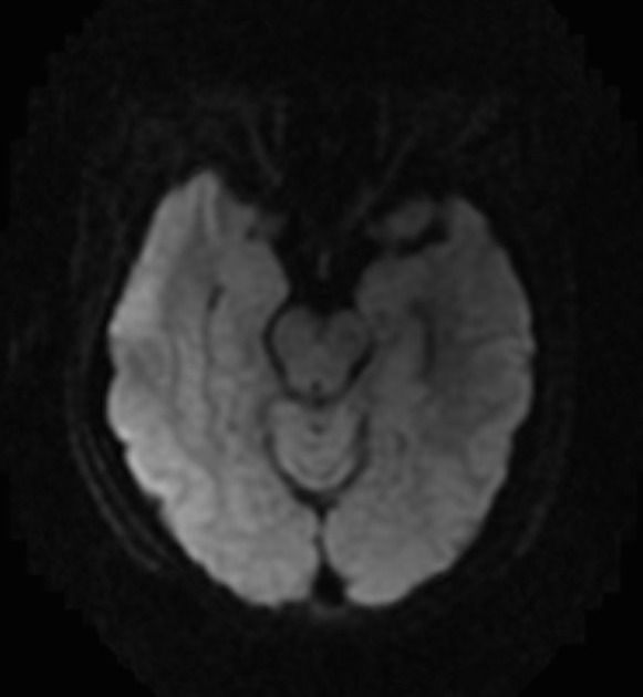
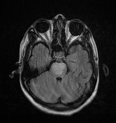
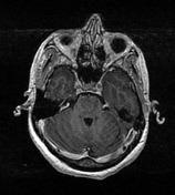
Brain MRI showed a heart-appearance area with restricted diffusion involving the anteromedial region of the mid-pons bilaterally. Subtle enhancement on T1 C+ of the restricted area is also noted. There is also right PICA infarction. The basilar artery appeared intact on MRI. Findings are compatible with acute/subacute infarct of the posterior circulation.
Case Discussion
This clinical picture is compatible with bilateral pontine infarct (posterior circulation). The heart-appearance sign is attributed to the involvement of bilateral paramedian and short circumferential pontine arteries.


 Unable to process the form. Check for errors and try again.