
Presentation
Evaluate renal abnormality.
Patient Data
Gender: Female
From the case:
Xanthogranulomatous pyelonephritis
Download
Info
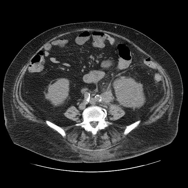
Enlarged left kidney with dilated calyces filled with low-attenuation material and a few small calcifications. Central staghorn calculus. Perinephric fluid/stranding with a small collection along the anterior upper pole. Ureteral thickening leading into an ill defined area of soft tissue stranding in the pelvis adjacent to a large cystic pelvic mass.
Case Discussion
Typical appearance of XGP which was treated with nephrectomy. During surgery, there were dense inflammatory adhesions surrounding the kidney and ureter, resulting in a very challenging surgery. The distal ureteral thickening was related to chronic inflammation and infection (and not the pelvic cyst).


 Unable to process the form. Check for errors and try again.