
Presentation
Head trauma. No loss of consciousness.
Patient Data


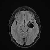
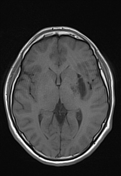
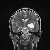
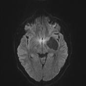
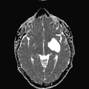
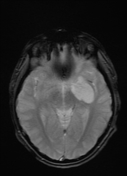
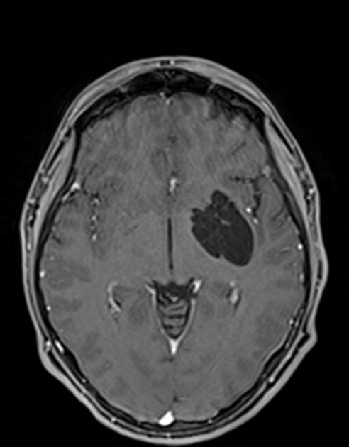
Intraparenchymal multiloculated cystic lesion of CSF intensity in the left basal ganglia extending to the left temporal lobe with minimal surrounding FLAIR hyperintensity. No restriction diffusion, enhancement or mass effect.
The lenticulostriate artery passes through the multiloculated cyst.
Case Discussion
Virchow-Robin spaces or perivascular spaces are small pial lined, cystic structures in the brain and are filled with interstitial fluid. They are normal spaces, identified in all age groups and are common in places where the penetrating vessels enter into the substance of the brain. Occasionally, these spaces can be enlarged and are termed as giant tumefactive perivascular spaces.1
It is important to recognize these to avoid unnecessary biopsy and potentially devastating complications.


 Unable to process the form. Check for errors and try again.