
Presentation
Postmenopausal bleeding
Patient Data


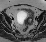
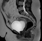
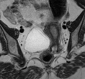
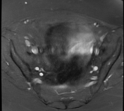
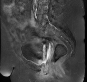
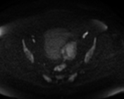
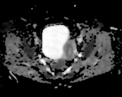
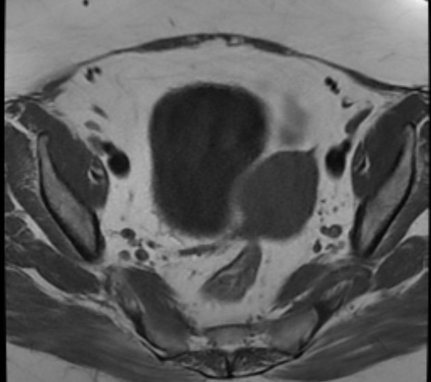
The MRI sequences demonstrate a pedunculated well-defined intracavitary mass arising from the fundal region, filling the uterine cavity and prolapsing through a distended endocervical canal. It appears isointense to the endometrium on T1, slightly hypointense on T2 with no restricted diffusion on DWI/ADC, surrounded by hyperintense fluid and endometrium with relatively homogeneous enhancement on postcontrast sequences. No infiltration of the myometrium. No pelvic lymphadenopathy or intraperitoneal effusion.
Case Discussion
MRI features are most consistent with an endometrial polyp.
On imaging, the main differential diagnosis is a pedunculated submucosal leiomyoma, which usually appears hypoechoic on ultrasound and demonstrates a hypointense signal on T2 MRI sequences.


 Unable to process the form. Check for errors and try again.