
Presentation
Presented with a headache. Blood workup showed low T4 and normal TSH.
Patient Data
Age: 13 years
Gender: Female
From the case:
Pituitary xanthogranuloma

Download
Info
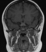
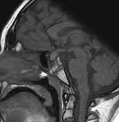
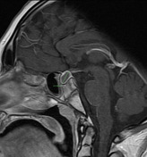
Rim-enhancing sellar mass with mild suprasellar bulging and stretching of the pituitary infundibulum. It has intermediate T1 and T2 signal centrally. There is compression of the optic chiasm.
Download
Info

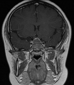
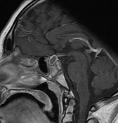
A single image from an MRI performed 3 months later showed further enlargement of the pituitary mass to 16 mm (from 13 mm).
Case Discussion
The sellar lesion increased in size on sequential imaging. It was then resected and pathology report confirmed pituitary xanthogranuloma.


 Unable to process the form. Check for errors and try again.