
Presentation
Dysphonia in an ex-smoker. Left submucosal lesion arising from left laryngeal ventricle on laryngoscopy.
Patient Data
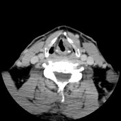

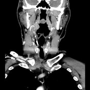

- left supraglottic laryngeal mass 1.6 x 1.5 x 1.2 cm centered above glottis, obliterating left laryngeal ventricle, displacing left aryepiglottic fold medially.
- no mass or suspicious asymmetry elsewhere within the upper aerodigestive tract
- no cervical lymphadenopathy

Intraoperative photo demonstrating left well-defined submucosal lesion arising from the left laryngeal ventricle, clear of vocal fold and anterior commissure.
Case Discussion
The patient underwent cold steel and shaver excision of the laryngeal lesion under general anesthesia.
Histology: numerous deposits of homogenous eosinophilic material which stained salmon pink on congo red and showed apple-green birefringence, confirming amyloidosis.
Case discussion
Amyloidosis is very rare and represents a heterogeneous group of disorders marked by abnormal protein formation and deposition which can cause organ failure and death. Up to 20% involve the head and neck, the larynx being the most common site of disease, representing 0.2-1.2% of all benign laryngeal tumors. Symptoms depend on the size and specific location of the deposit and may include dysphonia, dysphagia or exertional dyspnea.


 Unable to process the form. Check for errors and try again.